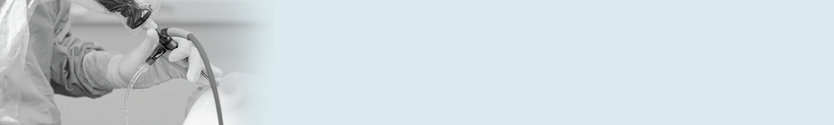


Arthroscopy of the Knee Joint
The arthroscope is a fibre-optic telescope that can be inserted into the knee joint to evaluate and treat a number of conditions. A camera is attached to the arthroscope and the picture is visualized on a high definition TV monitor. Most arthroscopic surgery of the knee is performed as ‘day case’ surgery and is usually done under general anesthesia. Knee arthroscopy is common, and millions of procedures are performed each year around the world.
Find out more about Arthroscopy of the Knee Joint with the following link
Arthroscopy is useful in evaluating and treating the following conditions
- Torn cartilage (meniscus): The cartilage is trimmed to a stable rim or repaired
- Torn surface (articular) cartilage
- Loose bodies within the joint (cartilage or bone that has broken off) and cysts
- Reconstruction of the Anterior Cruciate ligament/Posterior Cruciate Ligament
- Patello-femoral (knee-cap) disorders
- Washout of infected knees
- General diagnostic and planning purposes
Basic Knee Anatomy
The knee is one of the largest joints in the body. The knee joint is made up of the femur, tibia and patella (knee cap). All these bones are lined with articular (surface) cartilage. This articular cartilage acts like a shock absorber and provides a smooth, low friction surface for the knee to move on. Between the tibia and femur lie two cartilages called menisci. The medial (inner) meniscus and the lateral (outer) meniscus rest on the tibial surface cartilage and are mobile. The menisci also act as shock absorbers and stabilizers. The knee is stabilized by ligaments that are both in and outside the joint. The medial and lateral collateral ligaments support the knee from excessive side-to-side movement. The (internal) anterior and posterior cruciate ligaments stabilise the knee, preventing instability / giving way. The knee joint is surrounded by a capsule (envelope) that produces a small amount of synovial (lubrication) fluid to help with smooth motion. Thigh muscles are important secondary knee stabilizers.
Investigations
A routine X-Ray of the knee, which includes a standing weight-bearing view is usually required. An MRI scan which looks at the cartilages and soft tissues may be needed if the diagnosis is unclear. Occasionally, the use of Ultrasound or CT scan to investigate knee problems can be helpful.
Meniscal Cartilage Tears
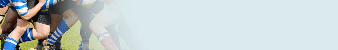
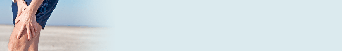
Following a twisting type of injury, the medial and lateral menisci can tear. This results either from a sporting injury or from a simple twisting injury such as getting out of a chair or standing from a squatting position. Our cartilages become a little brittle as we get older and therefore can tear a little more easily. The symptoms of a torn cartilage include
- Pain over the torn area i.e. inner or outer side of the knee
- Knee swelling
- Reduced motion
- Clicking, clunking or locking if the cartilage gets caught between the femur and tibia
Cartilage Tears
Once a meniscal cartilage has torn, it will not heal unless it is of a favourable pattern in an area with good blood supply (usually near the capsule of the joint). Once the cartilage has torn it no longer functions as a shock absorber, predisposing the knee to the risks of osteoarthritis (wear and tear) in later years. It is better to remove torn pieces from the knee if the knee is symptomatic.
Torn cartilages may continue to cause symptoms of mechanical discomfort, pain and swelling until the loose, ragged pieces are removed. Only the torn section is removed and the knee should recover and become symptom free. If the entire meniscus is removed, the knee may develop osteoarthritis in later years. It is standard to remove only the torn section of cartilage, preserving as much tissue as possible in an attempt to delay the onset of long-term ‘wear and tear’.
Provided the tear is of a favourable pattern (peripheral tear), in a young patient , the meniscus may be suitable for repair. If repaired, the patient must avoid twisting sports for a minimum of three months. A knee brace may also be required.
Articular Cartilage (Surface) Injury
If the surface cartilage is torn, this is significant, as a major shock-absorbing function is compromised. Large pieces of articular cartilage can ‘float’ in the knee (sometimes with bone attached) and this causes locking of the joint and can cause further deterioration due to the loose bodies unattached within the knee causing further damage. Most surface cartilage wear will ultimately lead to further osteoarthritis. Mechanical symptoms of pain and swelling due to cartilage peeling off can be helped with arthroscopic surgery. The surgery smoothes the edges of the surface cartilage and removes loose bodies.
Anterior Cruciate Ligament Injuries
Rupture of the Anterior (rarely the posterior) Cruciate Ligament (ACL) is a common sporting injury. Once ruptured the ACL does not heal and usually causes knee instability and the inability to return to normal sporting activities. An ACL reconstruction may be required and a new ligament is created to replace the ruptured ligament. This procedure is performed using the arthroscope.
Patella (knee-cap) Disorders
The arthroscope can be used to treat problems relating to kneecap disorders, particularly mal-tracking and significant surface cartilage tears. Patients may occasionally need to stay overnight if a lateral release has been performed as knee swelling is quite common. The majority of common kneecap problems can be treated with physical therapy and rehabilitation.
Inflammatory Arthritis
Occasionally arthroscopy is used in inflammatory conditions (e.g. Rheumatoid Arthritis) to help reduce the amount of inflamed synovium (joint lining) that is producing excess joint fluid. This procedure is called a synovectomy. After the surgery a drain may be inserted into the knee and the patient will generally require one or two nights in hospital.
Baker’s Cysts
Baker’s cysts or popliteal cysts are often found on clinical examination and ultrasound / MRI scan. The cyst is a fluid filled cavity behind the knee and in adults,may arise from a torn meniscus or worn articular cartilage in the knee. These cysts usually do not require removal as treating the cause (torn knee cartilage) will in most cases reduce the size of the cyst. Occasionally the cysts rupture and can cause calf pain. The cysts are not dangerous and do not require treatment if the knee is asymptomatic.
New Technology
Isolated areas of articular cartilage loss can be repaired using cartilage transplant technology. It is also possible to consider stem cell therapies which are showing some early promise. Further research is required to evaluate these options but it is a new and exciting field that is developing in the treatment of specific isolated cartilage defects in younger patients. Mr Edwards and Mr Melton are well placed to exploit these latest developments working closely with the Cambridge University Orthopaedic Research Department under the expert stewardship of Professor McCaskie.
The process is called Autologous Chondrocyte grafting . It involves harvesting cartilage cells from the affected knee, sending these cells to a laboratory and then culturing the cells to multiply into many cells. The large amount of cells produced are then placed back into the affected knee into the defect requiring resurfacing. Results are still short-term follow-up but appear to be encouraging.
After a major cartilage or ligament injury has been treated the knee can return to normal function. There is however a small increase in the risk of developing long-term wear and tear (Osteoarthritis) and depending on the degree of injury activity modification may be required. Activities that help prevent knees deteriorating quickly include:
- Low impact sports like swimming, cycling and walking
- Reducing weight and maintaining a healthy diet
Arthroscopy of the Knee: Patient Information
Please stop taking Aspirin and Anti-inflammatory medications 5 days prior to your surgery. You can continue taking all your other routine medication. If you smoke you are advised to stop prior to your surgery.
You will be admitted on the day of surgery and need to remain fasted for 6 hours prior to the procedure.
The limb undergoing the procedure will be marked and identified prior to the anesthetic being administered.
Once you are under an anesthetic, the knee is prepared in a sterile fashion. A tourniquet is placed around the thigh to allow a ‘blood – free’ procedure.
The arthroscope is introduced through a small (size of a pen) incision on the outer side of the knee. A second incision on the inner side of the knee is made to introduce the instruments that allow examination of the joint and treatment of the problem.
Post-operative Recovery
You will wake up in the recovery area and then be transferred back to the ward
A bandage will be around the operated knee.
Once you are recovered your intravenous drip will be removed and you will be shown a number of exercises to do.
Your surgeon will see you prior to discharge and explain the findings of the operation and what was done during surgery.
Pain medication will be provided and should be taken as directed
You can remove the bandage in 24-48 hours and retain the waterproof dressings over the wounds.
It is NORMAL for the knee to swell after the surgery. Elevating the leg when you are seated and placing ice packs on the knee will help to reduce swelling. (Ice packs on for 20 min 3-5 times a day until swelling has reduced)
You are able to return to work when comfortable unless otherwise instructed.
Please make an appointment 10-14 days after surgery to monitor your progress and remove any stitches in your knee.
Risks of Arthroscopy
General Anesthetic risks are extremely rare. Occasionally patients have some discomfort in the throat as a result of the tube that supplies oxygen and other gasses. Please discuss with the Specialist Anesthetist if you have any specific concerns
Risks related to Arthroscopic Knee Surgery Include
- Postoperative bleeding
- Deep Vein Thrombosis
- Infection
- Stiffness
- Numbness to part of the skin near the incisions
- Injury to vessels, nerves and chronic pain syndrome
- Progression of the disease process
The risks and complications of arthroscopic knee surgery are extremely small. One must however bear in mind that occasionally there is more damage in the knee than was initially thought and that this may affect the recovery time. In addition, if the cartilage in the knee is partly worn out then arthroscopic surgery has about a 65% chance of improving symptoms in the short to medium term but more definitive surgery may be required in the future. In general arthroscopic surgery does not improve knees that have well established osteoarthritis.
Post-Operative Exercises and Physical Therapy
Following your surgery, you will be given an instruction sheet showing exercises that are helpful in speeding up your recovery. Strengthening your thigh muscles (Quadriceps and Hamstrings) is most important. Swimming and cycling (stationary or road) are excellent ways to build these muscles up and improve movement.
Frequently asked questions
How long am I in the Hospital?
A: Between 4-6 hours
Do I need crutches?
A: Usually not required (Unless you are having Anterior Cruciate Ligament Reconstruction)
When can I get the knee wet?
A: After 24 hrs remove the bandage and retain waterproof dressings.
When can I drive?
A: After 48 hrs if the knee is comfortable. You need to assess if you are able to do an emergency stop without pain
When can I return to work?
A: When the knee feels reasonably comfortable.
When can I shower?
A: After 24 hrs provided the dressings/wounds can be kept completely dry
When can I swim or bathe?
A: After removal of the stitches.
How long will my knee take to recover?
A: Depending on the findings and surgery, usually 4 to 6 weeks following the surgery.
When Can I return to Sports?
A: Depending on the findings, 4-6 weeks after surgery.